Pictures from what we do
![](https://ingestem.org/wp-content/uploads/2015/09/Colonies-de-cellules-iPS-humaines-bandeau-021-300x115.jpg)
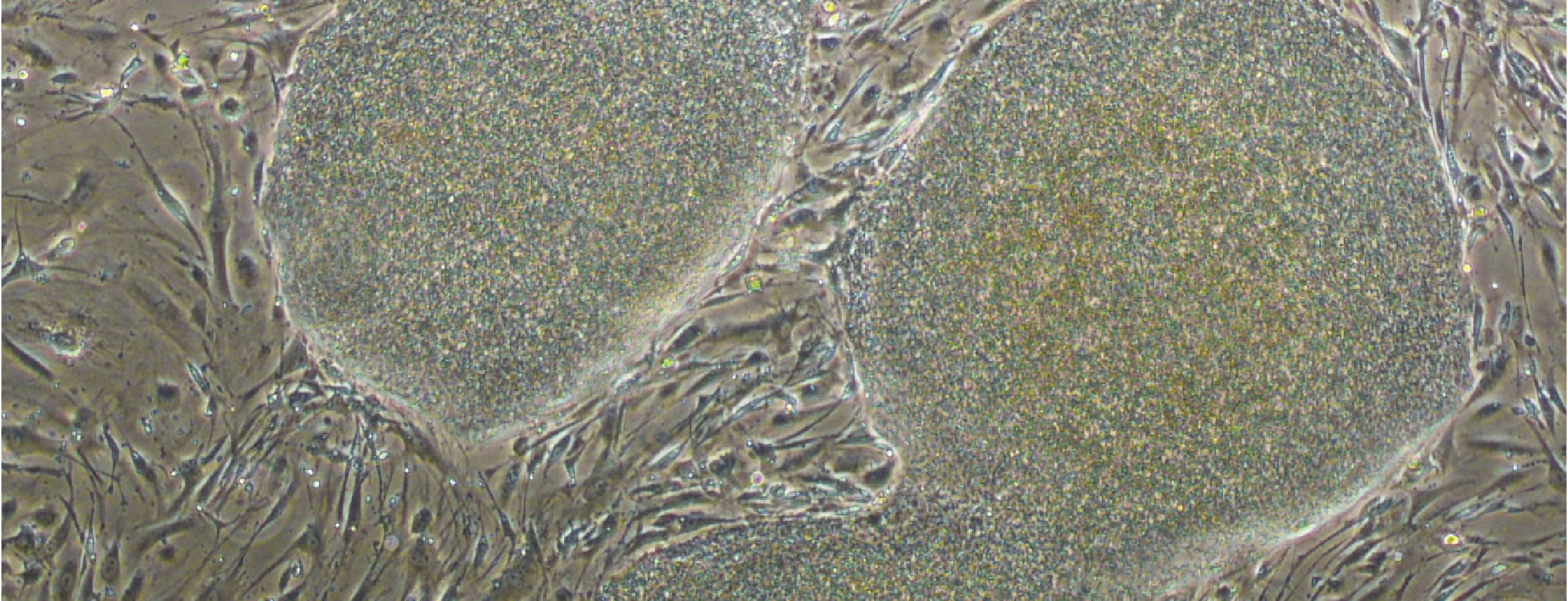
Human iPS cell colonies
![](https://ingestem.org/wp-content/uploads/2015/09/Epid_hESC_rec1-300x144.png)
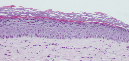
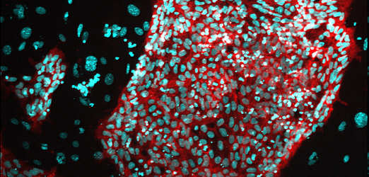
Epithelium formed from human embryonic stem cells. © AFMTELETHON / Luc Morvan
![](https://ingestem.org/wp-content/uploads/2015/09/28164556-1-300x144.png)
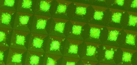
Fluorescent iPS cell colonies in microwell plates Aggrewell StemCell Technologies. © ESTEAM Paris Sud
![](https://ingestem.org/wp-content/uploads/2015/09/MG_4432-300x144.png)
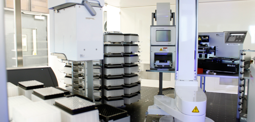
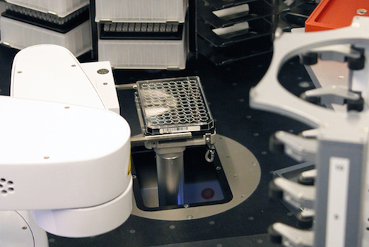
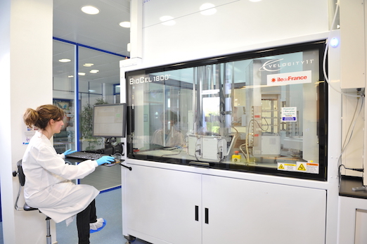
Cellular high throughput screening platform. Biocel System - Agilent Technologies. © IGBMC
![](https://ingestem.org/wp-content/uploads/2015/09/INGESTEM-Colonie-cellules-ES-macaque-300x144.png)
Colony of Macaque ES cells ©PrimaStem
![](https://ingestem.org/wp-content/uploads/2015/04/DSC_2652-300x199.jpg)
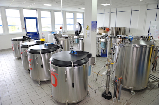
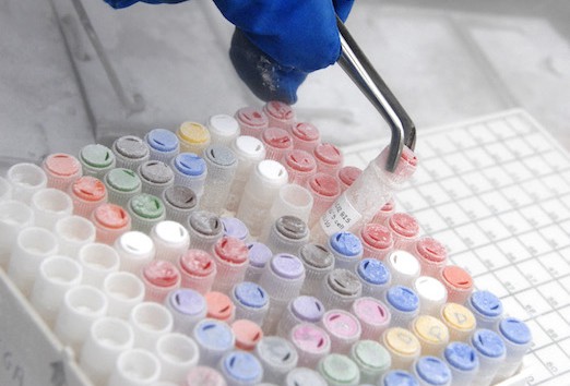
Cryogenic storage of cell lines bank © AFMTELETHON / Luc Morvan
![](https://ingestem.org/wp-content/uploads/2015/09/Aggregat_neuron_hESC_DM1_reso1-300x144.png)
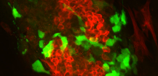
Aggregates of neurons carrying the mutation DM1 (Myotonic Dystrophy Type I) differentiated from human embryonic stem cells © I-Stem
![](https://ingestem.org/wp-content/uploads/2015/09/image_0734-300x144.png)
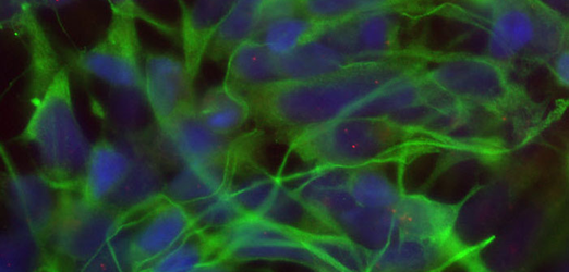
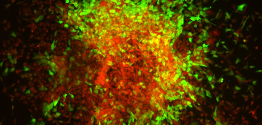
Colony of murine differentiated embryonic stem cell, expressing a reporter of paraxial mesoderm (green), and a neural protein (red). © IGBMC
![](https://ingestem.org/wp-content/uploads/2015/04/HTS5-03-300x201.jpg)
![](https://ingestem.org/wp-content/uploads/2015/09/INGESTEM-Cellules-en-cours-de-differenciation-avec-gene-rapporteur-300x144.png)
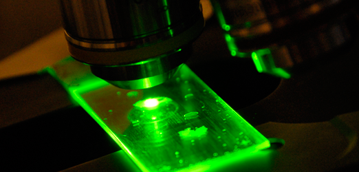
Cells undergoing differentiation with reporter gene
![](https://ingestem.org/wp-content/uploads/2015/04/ajp71046-e1442313361150-300x203.jpg)
![](https://ingestem.org/wp-content/uploads/2015/04/HTS2-300x200.jpg)
![](https://ingestem.org/wp-content/uploads/2015/04/HTS1-300x199.jpg)
![](https://ingestem.org/wp-content/uploads/2015/07/ajp70791-300x144.png)
ajp70791
![](https://ingestem.org/wp-content/uploads/2015/04/MG_4432-300x144.png)